Anatomy Of Chest And Stomach / Abdominal and Pelvic CT / There are two sphincters of the stomach, located at each orifice.. This page provides an overview of the chest muscle group. Its size and shape changes from time to time depending on the volume of its contents (food/fluid). The thoracic cage is the bony. Anatomy of the chest and the lungs: The upper portion of the trunk between the neck and the abdomen.
Swensen fund for innovation in teaching. They control the passage of material entering and a hiatus hernia occurs when a part of the stomach protrudes into the chest through the oesophageal. When having a discussion concerning anatomy the phrase form determines function comes to mind. This type of ct scan uses a lower radiation level than a conventional. The thoracic cage is the bony.
Internal Anatomy Of Male Chest And Abdomen On Black Stock ... from media.istockphoto.com The embryologic and anatomic basis of modern surgery. Layers serosa or visceral peritoneum: Between oesophagus (ostium cardiacum) and small intestine (ostium pyloricum) syntopy (syntopia): Find out more about the individual muscles within the chest anatomy by clicking their respective links throughout this page. The stomach lies within the superior aspect of the abdomen. The thoracic cage is the bony. To duodenum regions cardiac fundus body pyloric. It lies between the chest and the pelvis, holding many of the body's organs.
It lies between the chest and the pelvis, holding many of the body's organs.
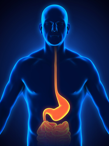
The stomach consists of several important anatomical parts. Grant jcb, basmajian jv, slonecker ce. The anatomical drawings were organized in a fairly classical manner to be easily used as a standard anatomical atlas. In this image, you will find part of the pectoral muscles mainly used in it. Learn about the anatomy of the stomach and the types of cells that cover the stomach. Ingested food enters the stomach from the esophagus via the cardiac orifice, falling into gastric juice produced by the stomach. Between oesophagus (ostium cardiacum) and small intestine (ostium pyloricum) syntopy (syntopia): It's part of the digestive system. The chest wall is supplied by the posterior intercostal arteries arising from the aorta, the internal thoracic and the highest intercostals given off the subclavian artery, and the branches of the axillary artery (fig. It is part of the digestive system, which extends from the mouth to the anus. There are two sphincters of the stomach, located at each orifice. Lee mcgregor's synopsis of surgcial anatomy. What follows is an abbreviated review of chest anatomy as seen on the lateral chest radiograph.
This video is about the division of the abdominal cavity and the anatomy of the stomach. Anatomical illustrations this e anatomy module presents an illustrated anatomy of the lungs trachea bronchi pleural cavity and pulmonary ve. The stomach consists of several important anatomical parts. Protective framework for parts of the chest involved with brea… chest or thorax. This type of ct scan uses a lower radiation level than a conventional.
Human Stomach Anatomy Stock Photo - Download Image Now ... from media.istockphoto.com Layers serosa or visceral peritoneum: It's part of the digestive system. The chest wall is supplied by the posterior intercostal arteries arising from the aorta, the internal thoracic and the highest intercostals given off the subclavian artery, and the branches of the axillary artery (fig. Ingested food enters the stomach from the esophagus via the cardiac orifice, falling into gastric juice produced by the stomach. The stomach consists of several important anatomical parts. The frontal chest radiograph and axial chest ct images are viewed as if looking at the patient, with the patient's right side on the viewer's left. Grant jcb, basmajian jv, slonecker ce. The stomach is a muscular, hollow organ in the gastrointestinal tract of humans and many other animals, including several invertebrates.
The anatomical drawings were organized in a fairly classical manner to be easily used as a standard anatomical atlas.
The stomach lies within the superior aspect of the abdomen. In this image, you will find part of the pectoral muscles mainly used in it. This page provides an overview of the chest muscle group. The thoracic cage is the bony. Between oesophagus (ostium cardiacum) and small intestine (ostium pyloricum) syntopy (syntopia): You may also find triceps, lateral head brachialis, biceps brachii, latissimus dorsi, deltoid, acromion anatomynote.com found chest muscle anatomy from plenty of anatomical pictures on the internet. Anatomy of the chest and the lungs: The groin is the area in the body where the upper thighs meet the lowest part of the abdomen. This type of ct scan uses a lower radiation level than a conventional. Stomach blood supply arterial blood supply: Stimulated by the acidic condition in the stomach and certain other factors, these cells release pepsin enzyme in its inactive form, called pepsinogen, which then carries out the digestion of proteins. Learn about the anatomy and physiology of the stomach. In the anatomy of stomach, it is the uppermost portion, forming the upper curvature of the organ.
Learn about the anatomy of the stomach and the types of cells that cover the stomach. The upper portion of the trunk between the neck and the abdomen. You may also find triceps, lateral head brachialis, biceps brachii, latissimus dorsi, deltoid, acromion anatomynote.com found chest muscle anatomy from plenty of anatomical pictures on the internet. Anatomy of the stomach, gallbladder, and pancreas. This page provides an overview of the chest muscle group.
Vintage 1950's Frohse Chest & Abdomen Viscera Human ... from cdn.shopify.com Stomach blood supply arterial blood supply: What this entails is that the structure of the organ determines strongly the stomach is an expanded section of the gastrointestinal tract between the esophagus and the duodenum of the small intestine. The thoracic cage is the bony. The embryologic and anatomic basis of modern surgery. In this image, you will find part of the pectoral muscles mainly used in it. Stimulated by the acidic condition in the stomach and certain other factors, these cells release pepsin enzyme in its inactive form, called pepsinogen, which then carries out the digestion of proteins. Swensen fund for innovation in teaching. In the anatomy of stomach, it is the uppermost portion, forming the upper curvature of the organ.
Grant jcb, basmajian jv, slonecker ce.
It is part of the digestive system, which extends from the mouth to the anus. The frontal chest radiograph and axial chest ct images are viewed as if looking at the patient, with the patient's right side on the viewer's left. In the anatomy of stomach, it is the uppermost portion, forming the upper curvature of the organ. Gross anatomy the stomach is a rounded, hollow organ located just inferior to the diaphragm in the left part of the abdominal cavity. The thoracic cage is the bony. The chest anatomy includes the pectoralis major, pectoralis minor and the serratus anterior. Although there is no anatomical sphincter to be demonstrated at the lower end of the oesophagus in man, a multifactorial 'physiological'sphincter mechanism is the oesophagus extends from the distal termination of the pharynx at the level of the sixth cervical vertebra to the cardiac orice of the stomach. Swensen fund for innovation in teaching. Learn about the anatomy of the stomach and the types of cells that cover the stomach. The stomach is much like a bag with a lining. The stomach is a muscular, hollow organ in the gastrointestinal tract of humans and many other animals, including several invertebrates. The stomach receives food from the esophagus. Grant jcb, basmajian jv, slonecker ce.
Learn more from webmd about the anatomy of the stomach, along with illnesses that affect the stomach and tests to diagnose stomach problems anatomy of chest. False flatter chest fluctuating fluoride.


Posting Komentar
0 Komentar